Morphology of Leydig cells in the testes after in vivo MCP-1 treatment.
Por um escritor misterioso
Descrição

Morphology of Leydig cells in the testes after in vivo PTHrP
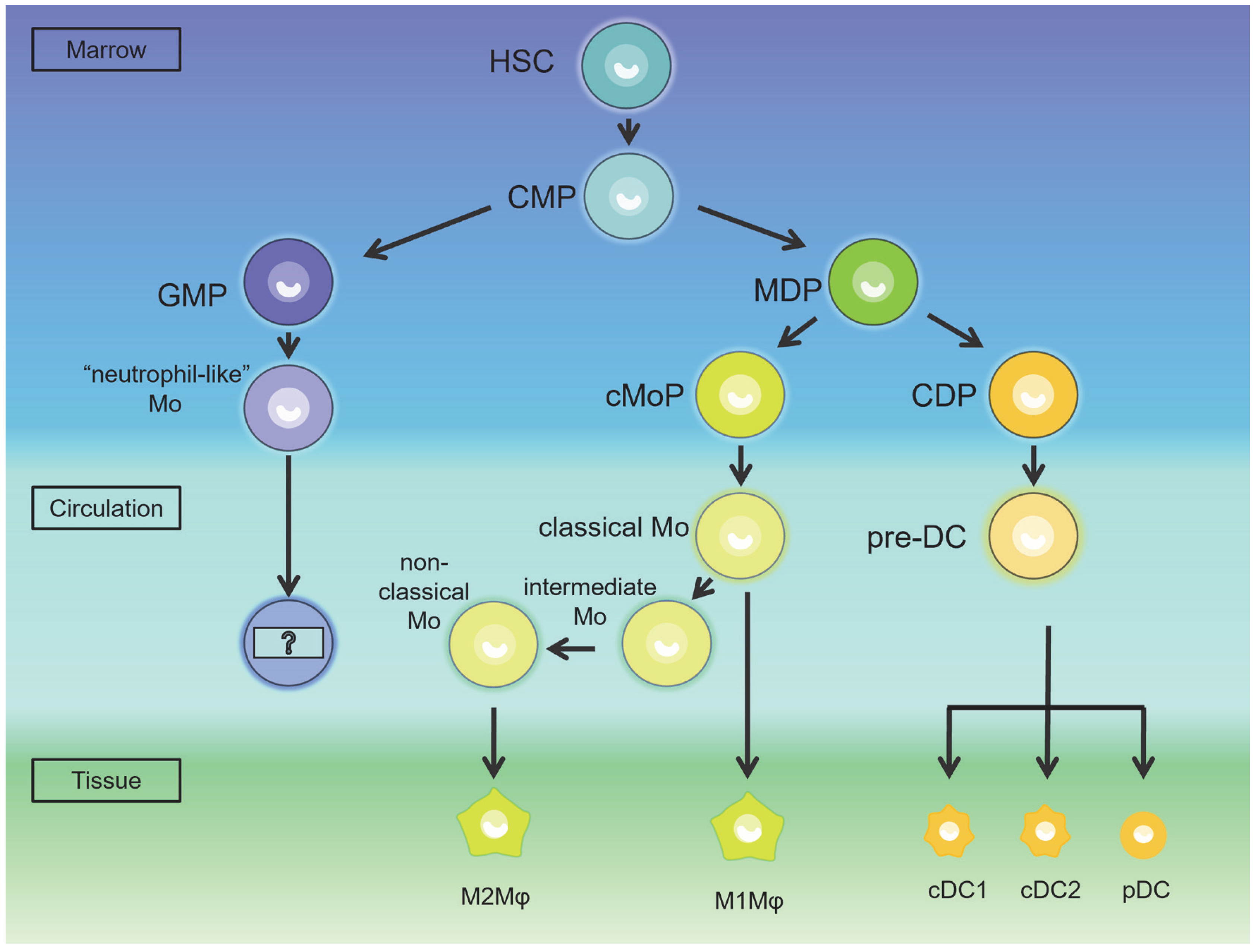
IJMS, Free Full-Text

Stem Leydig cells: Current research and future prospects of regenerative medicine of male reproductive health - ScienceDirect

Testicular torsion in vivo models: Mechanisms and treatments - Minas - 2023 - Andrology - Wiley Online Library
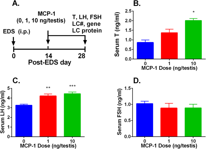
Monocyte Chemoattractant Protein-1 stimulates the differentiation of rat stem and progenitor Leydig cells during regeneration, BMC Developmental Biology

Testicular macrophages are recruited during a narrow time window by fetal Sertoli cells to promote organ-specific developmental functions

Morphology of Leydig cells in the testes after in vivo MCP-1 treatment.
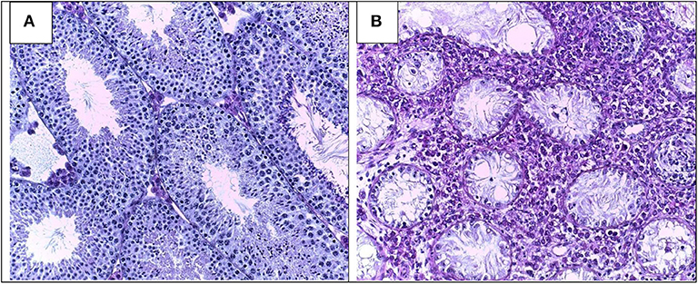
Frontiers Pathomechanisms of Autoimmune Based Testicular Inflammation
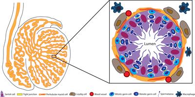
Frontiers Sertoli Cell Immune Regulation: A Double-Edged Sword
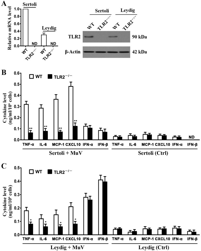
Mumps virus-induced innate immune responses in mouse Sertoli and Leydig cells

Characterization of the structural, oxidative, and immunological features of testis tissue from Zucker diabetic fatty rats
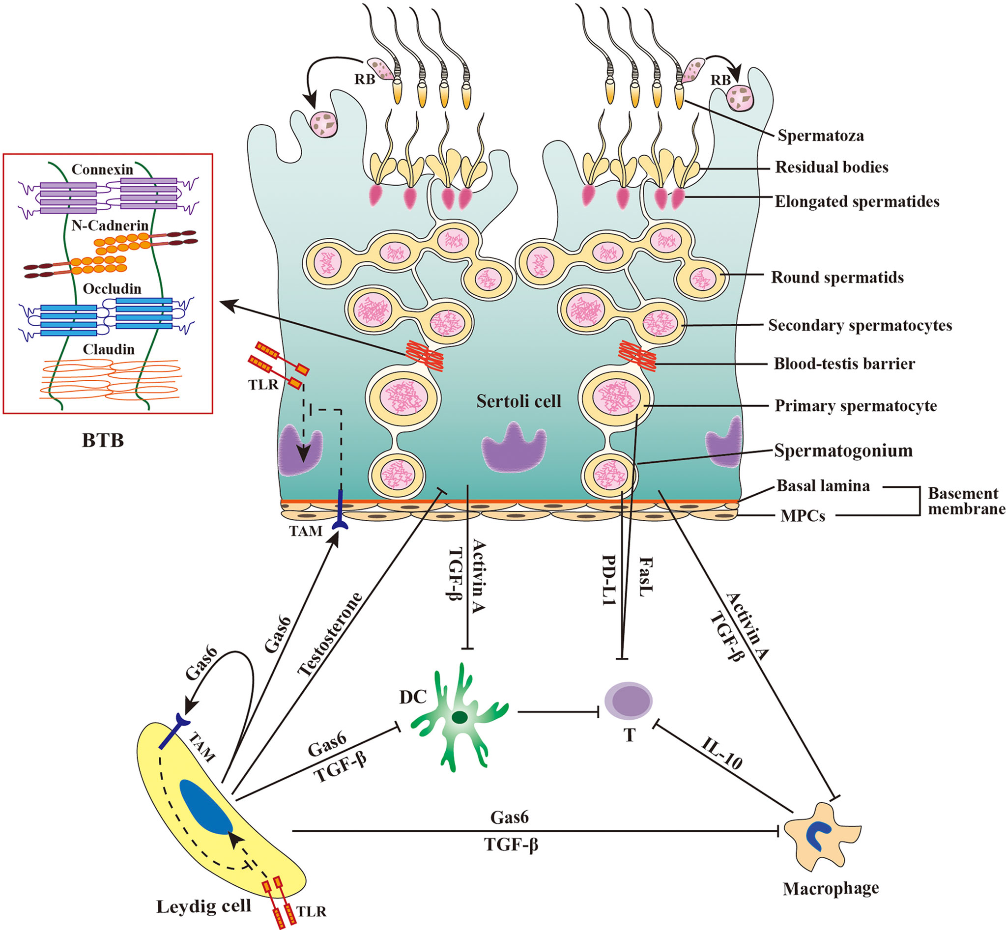
Frontiers Viral tropism for the testis and sexual transmission
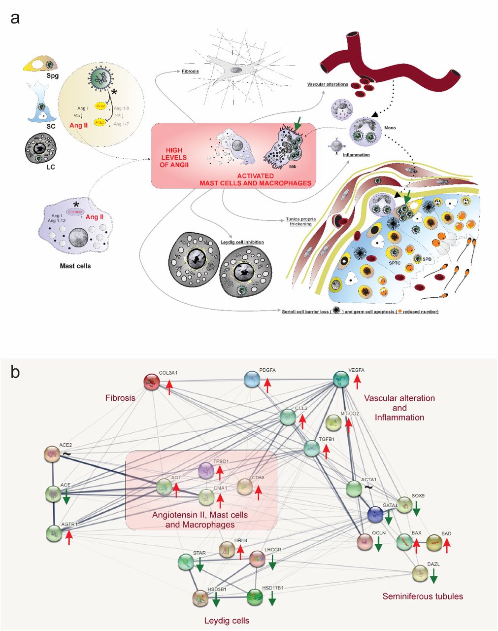
SARS-CoV-2 infects, replicates, elevates angiotensin II and activates immune cells in human testes
de
por adulto (o preço varia de acordo com o tamanho do grupo)
