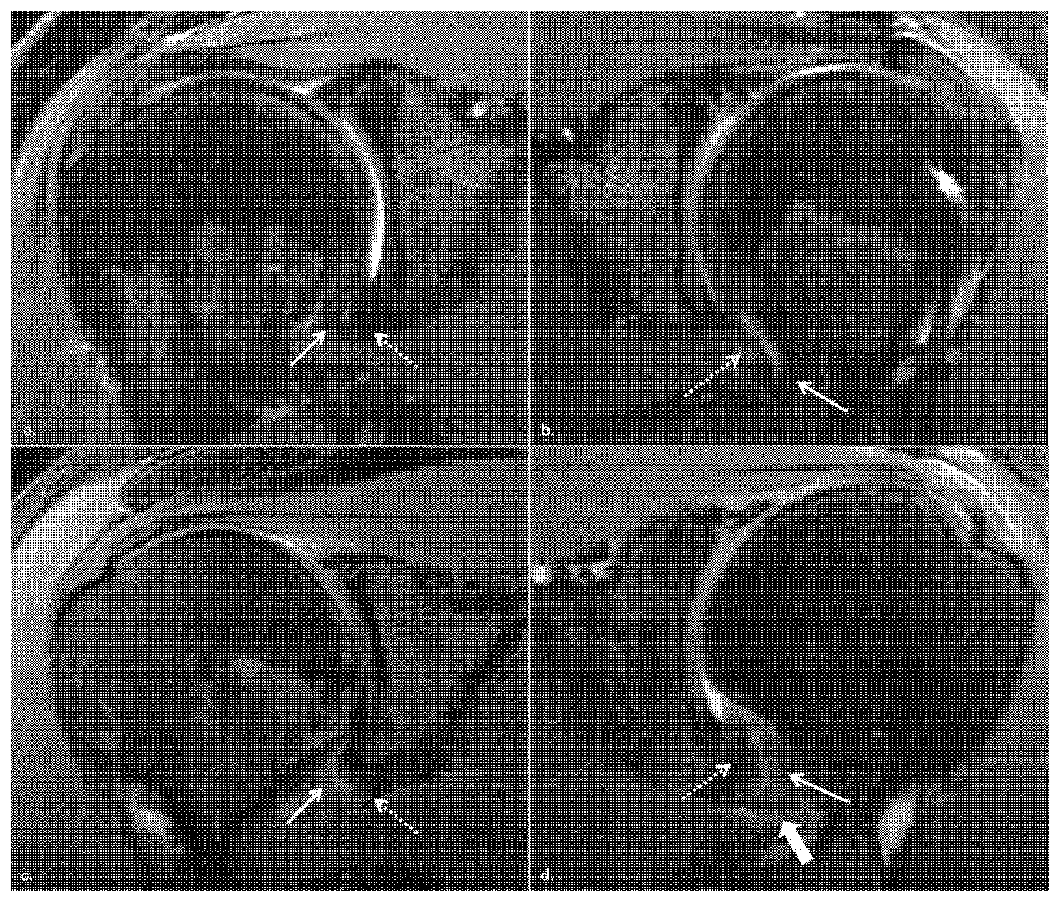
Typical magnetic resonance imaging scan showing the coracohumeral
Por um escritor misterioso
Descrição

The Rotator Interval: A Review of Anatomy, Function, and Normal and Abnormal MRI Appearance

Imaging of the Acromioclavicular Joint: Anatomy, Function, Pathologic Features, and Treatment

Imaging of Tendons

BIR Publications

A 61-year-old man with adhesive capsulitis of the shoulder. (AeC) MRI

Beyond the Cuff: MR Imaging of Labroligamentous Injuries in the Athletic Shoulder
:background_color(FFFFFF):format(jpeg)/images/library/13522/Shoulder_joint.png)
Normal shoulder MRI: How to read a shoulder MRI

Magnetic resonance imaging of the shoulder

JCM, Free Full-Text
de
por adulto (o preço varia de acordo com o tamanho do grupo)


